MRIndex Module - muscle MRI (mMRI)
The mMRI (muscle imaging methodology) has represented for years an important non-invasive and reliable definition tool for the clinical diagnosis and follow-up of MNM and can already be used as a possible biomarker in pharmacological clinical trial studies. In fact, MRI can detect not only the compactness of the individual muscle belly in individual body areas but also the presence of adipose tissue within the muscle, allowing the staging of tissue damage and its correlation with the progression of the clinical picture. This finding is in fact a marker linked to the severity and progression of the pathology and can therefore provide useful information to the clinician regarding its course. At the moment, however, the evaluation of muscular impairment with imaging occurs mostly through visual gestalt by a few experts in the field, representing a subjective and qualitative method, where arbitration criteria linked to the skills and experience of the operator always weigh more, especially in the early phase of the disease and in clinical pictures that are difficult to genetically define. This is partly due to the fact that there is still no quantitative analysis software for muscle imaging and this creates inevitable repercussions on diagnostic effectiveness and accuracy. It is conceivable that standardization procedures and the use of quantitative methods can optimize diagnostic performance. In the past, several attempts have been made to extract an objective score regarding this factor, including the analysis of T1-weighted magnetic resonance images after segmentation. Since then, several improvements have been made to the algorithm, which in its current version was presented at an international conference, and is now integrated into the dedicated MRIndex module of the Health360 platform developed for the InGene project.
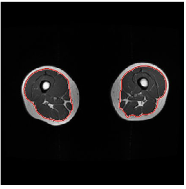
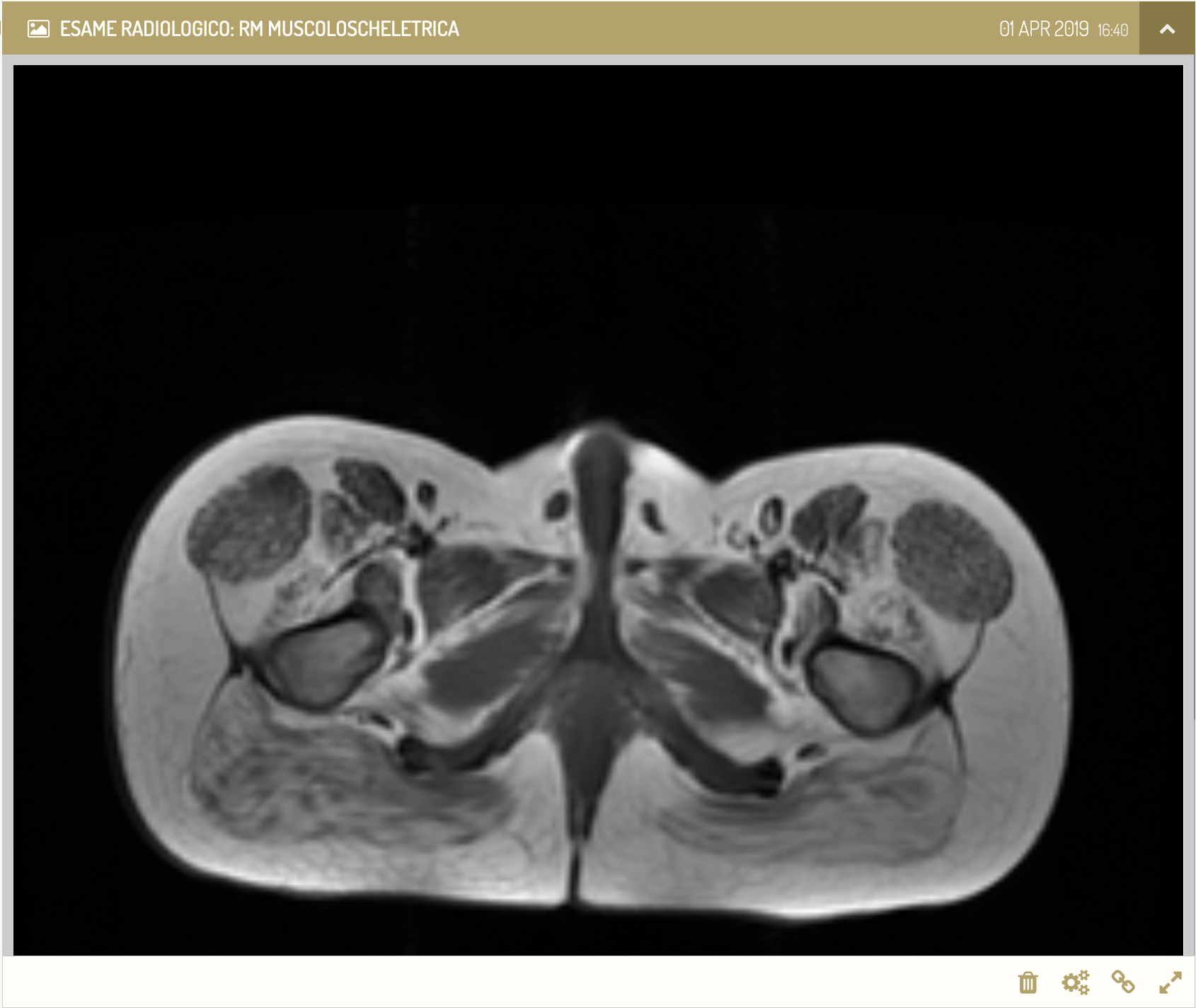
The objective of Muscle Magnetic Resonance Imaging is therefore to evaluate how much muscle function is compromised. This evaluation, which is expressed through an index varying between 1 and 5, is obtained by evaluating the percentage of "white" in the muscle imaged via MRI. The white part in fact reflects the fatty component of the limb, the infiltration of which into the muscular part increases with the decrease in the functionality of the muscle and consequently its mass. The development of MRIndex involved, as a fundamental step, the use of Orthanc (RIS-PACS server on which the DICOM files relating to the various exams are uploaded) for the relevant module. The feature is visible as a module in the menu part and as an event in the timeline of the individual patient after loading the radiological exam from the menu in the timeline itself. Only for those exams in which the application of the aforementioned algorithm is possible, a button will appear in the event through which to trigger the calculation and, once processing has taken place by the algorithm, the user will be presented with a new event in the timeline with the result obtained (percentage of fat present) and the possibility of comparing it with a possible previous sending through specific graphs.
Specifically, in the patient's timeline it is possible to view the test to which the patient has undergone. By pressing the first button on the left under the image, an application will be launched in the background, integrated into the H360 platform, which will calculate the adipose infiltration in the muscle tissue of the area considered. The application will therefore use the algorithms developed by the group of the Department of Physics of the University of Pisa involved in the InGene project thanks to the close and multi-year collaboration with IRCCS Fondazione Stella Maris. Once the algorithm, after appropriate segmentation of the image, has finished its work, a new event will be created on the patient's timeline with the detail of the result obtained.
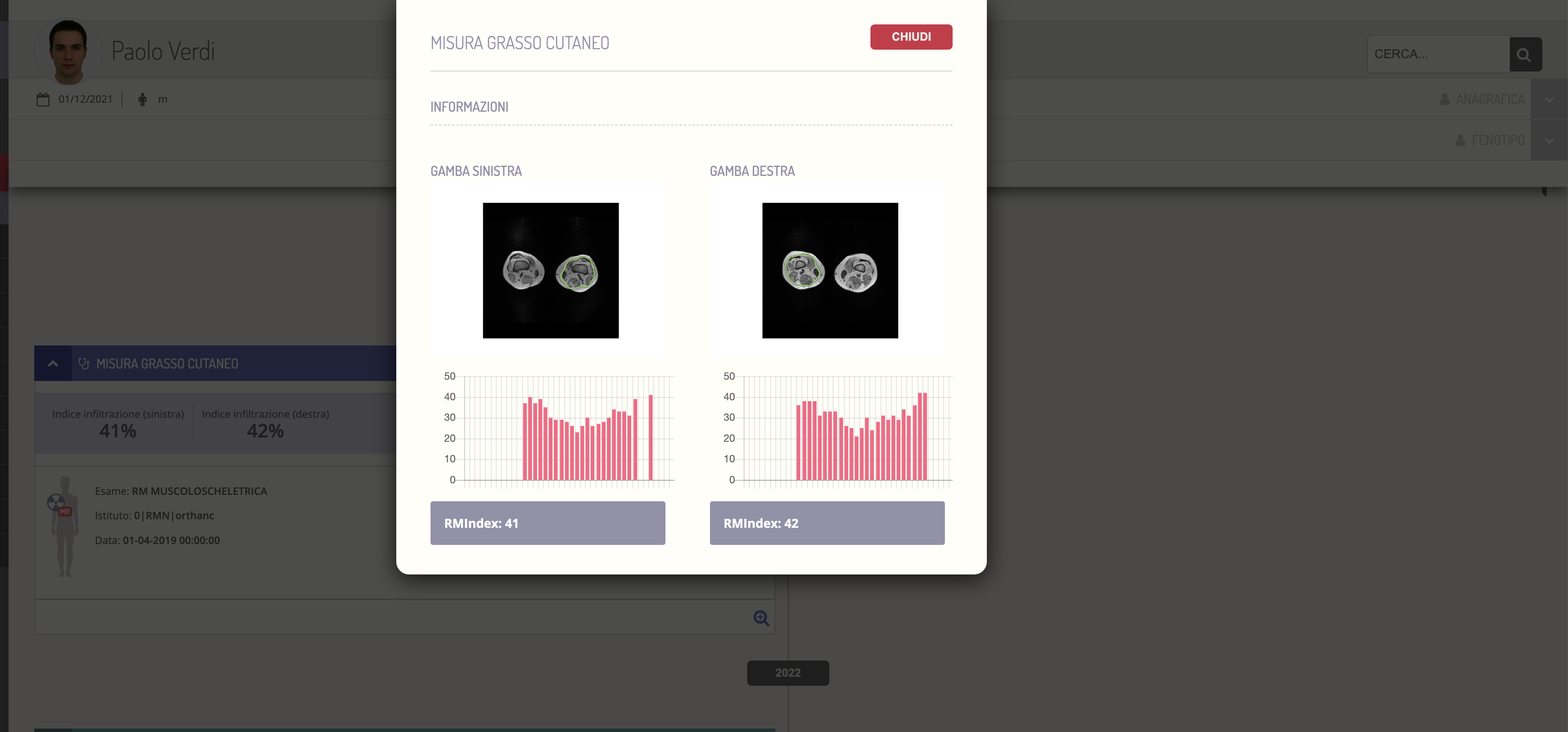
The final result can then be displayed both numerically and graphically, in relation to each of the two limbs, as well as to each slice possibly loaded, in such a way as to make it possible for the clinician to calculate the RMIndex along different segments of the lower limb.